Hi, I'm Ruben van der Waerden
A passionate PhD candidate at Radboudumc (Nijmegen) with expertise in artificial intelligence for medical imaging.
About Me
My Story
Ruben van der Waerden (1997) is a PhD candidate at the CARA Lab as part of the Diagnostic Image Analysis Group. He completed both his Bachelor’s Degree in Biomedical Engineering and Master’s Degree in Medical Engineering at the Technical University of Eindhoven.
During his master’s studies, Ruben developed a keen interest in AI applications within the medical field. His master’s thesis focused on deriving blood pressure from non-invasive photoplethysmogram (PPG) signals using deep learning models. Since March 2023, Ruben has been working as a PhD candidate within the Cardiology department. His research focuses on automated analysis of optical coherence tomography (OCT) images during cardiac catheterization procedures, aiming to develop a new method for assessing the risk of myocardial infarction in patients with coronary artery disease. This research is supervised by Niels van Royen, Ivana Isgum, and Jos Thannhauser.
My Skills
Python
Docker
Slurm
C
Git
Experience
PhD Candidate
CARA Lab (Radboudumc)
Development of automatic segmentation of intravascular optical coherence tomography (IVOCT) using deep learning.
Intern Researcher
The University of Sheffield
Development of a mathematical and physiological model of a human vein, using Lattice Boltzmann modelling
Intern Researcher
FreeSense Solutions
AI-derived blood pressure estimation based on photoplethysmogram (PPG).
My Articles
These are my highlighted articles. For all articles, visit my ResearchGate profile!
Comprehensive full-vessel segmentation and volumetric plaque quantification for intracoronary optical coherence tomography using deep learning
Rick Volleberg, Ruben van der Waerden, Niels van Royen
Intracoronary optical coherence tomography (OCT) provides detailed information on coronary lesions, but interpretation of OCT images is time-consuming and subject to interobserver variability. The aim of this study was to develop and validate a deep learning-based multiclass semantic segmentation algorithm for OCT (OCT-AID). A reference standard was obtained through manual multiclass annotation (guidewire artefact, lumen, side branch, intima, media, lipid plaque, calcified plaque, thrombus, plaque rupture, and background) of OCT images from a representative subset of pullbacks from the PECTUS-obs study. Pullbacks were randomly divided into a training and internal test set. An additional independent dataset was used for external testing. In total, 2808 frames were used for training and 218 for internal testing. The external test set comprised 392 frames. On the internal test set, the mean Dice score across nine classes was 0.659 overall and 0.757 on the true-positive frames, ranging from 0.281 to 0.989 per class. Substantial to almost perfect agreement was achieved for frame-wise identification of both lipid (κ=0.817, 95% CI 0.743–0.891) and calcified plaques (κ=0.795, 95% CI 0.703–0.887). For plaque quantification (e.g. lipid arc, calcium thickness), intraclass correlations of 0.664–0.884 were achieved. In the external test set, κ-values for lipid and calcified plaques were 0.720 (95% CI 0.640–0.800) and 0.851 (95% CI 0.794–0.908), respectively. The developed multiclass semantic segmentation method for intracoronary OCT images demonstrated promising capabilities for various classes, while having included difficult frames, such as those containing artefacts or destabilized plaques. This algorithm is an important step towards comprehensive and standardized OCT image interpretation.
Click here for full text!Artificial intelligence for the analysis of intracoronary optical coherence tomography images: a systematic review
Ruben van der Waerden, Rick Volleberg, Jos Thannhauser
Intracoronary optical coherence tomography (OCT) is a valuable tool for, among others, periprocedural guidance of percutaneous coronary revascularization and the assessment of stent failure. However, manual OCT image interpretation is challenging and time-consuming, which limits widespread clinical adoption. Automated analysis of OCT frames using artificial intelligence (AI) offers a potential solution. For example, AI can be employed for automated OCT image interpretation, plaque quantification, and clinical event prediction. Many AI models for these purposes have been proposed in recent years. However, these models have not been systematically evaluated in terms of model characteristics, performances, and bias. We performed a systematic review of AI models developed for OCT analysis to evaluate the trends and performances, including a systematic evaluation of potential sources of bias in model development and evaluation.
Click here for full text!Analysis of Blood Stasis for Stent Thrombosis Using an Advection-Diffusion Lattice Boltzmann Scheme
Ruben van der Waerden, Ian Halliday
An advection-diffusion solver was applied to assess how stent strut shape and position impact the development of a pro-thrombotic region within the stented human artery. Presented here is a suitably parameterised advection-diffusion equation with a source term that is spatially uniform within a certain sub-domain of interest to compute a “time concentration”. The latter will serve as a surrogate quantity for the “age” of fluid parcels, i.e., the time the fluid parcel has spent in the sub-domain. This is a particularly useful concept in the context of coronary artery haemodynamics, where “stasis of blood” (or residence time) is recognized as the most important factor in thrombotic initiation. The novel method presented in this work has a very straightforward and convenient single lattice Boltzmann simulation framework encapsulation. A residence time surrogate is computed, presented and correlated with a range of traditional haemodynamic metrics (wall shear stress, shear rate and re-circulation region shapes) and finally, the role of these data to quantify the risk of thrombus formation is assessed.
Click here for full text!Press and Conferences
2025
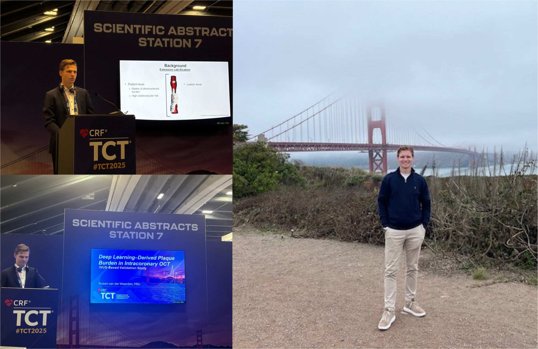
TCT Congress San Francisco
I presented our work on AI-based calcium volume assessment using OCT, focusing on its interaction with plaque vulnerability features in a lesion-level analysis. This study is currently in submission phase as a full manuscript. In addition, I presented research I contributed to on OCT-based plaque burden quantification using our AI software for complete vessel segmentation, published as a research letter inJACC: Cardiovascular Interventions. The TCT Congress was an inspiring event with many insightful sessions, and some time to explore San Francisco, including cycling across the Golden Gate Bridge.
Featured in Dutch newspapers, and on live TV
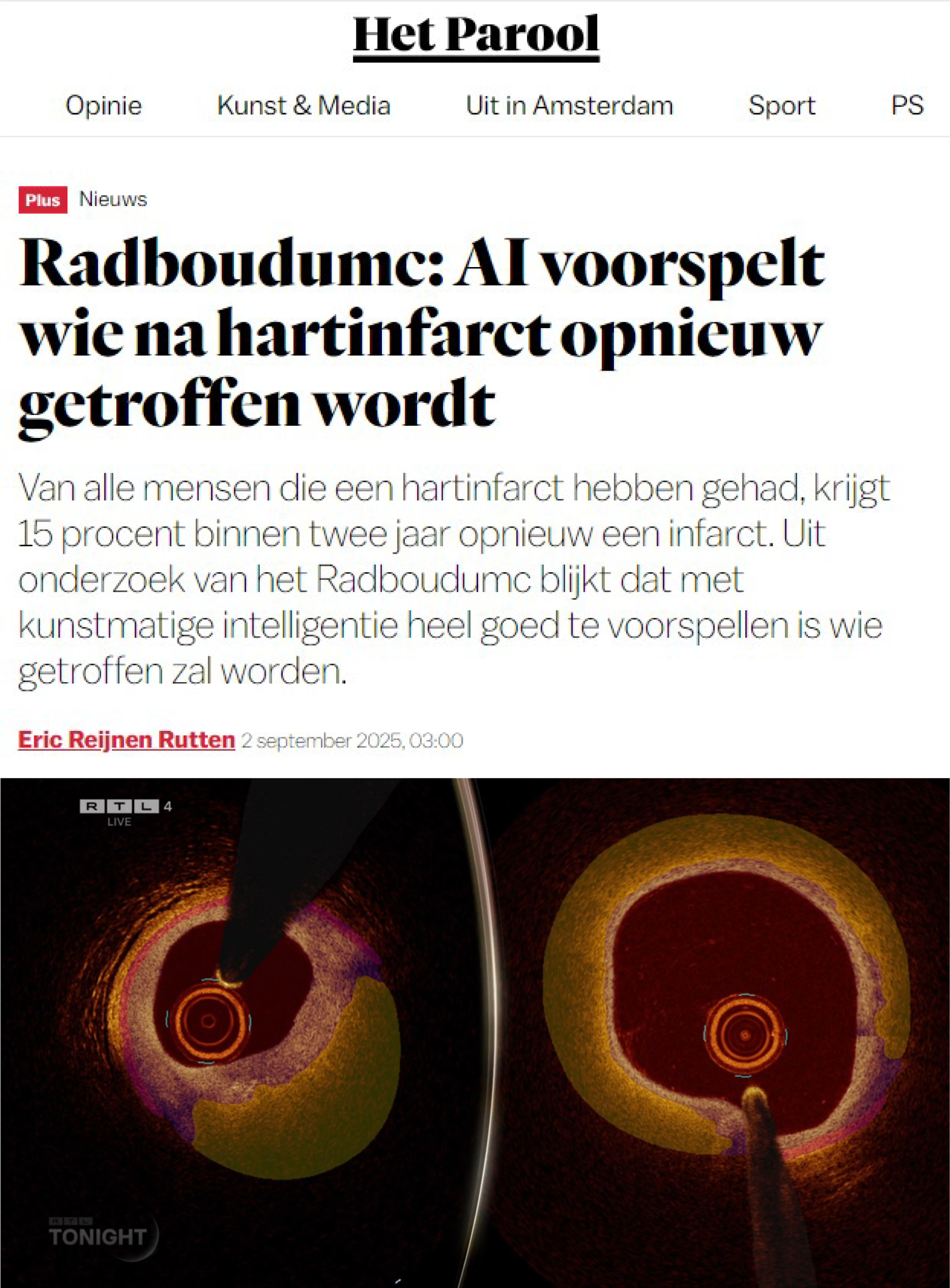
Our research was presented at the ESC Congress on 01-09-2025. The study evaluated the utility of artificial intelligence (AI) for identifying thin-cap fibroatheroma (TCFA) in relation to clinical outcomes. As part of my PhD work, I focus on developing pixelwise segmentation models, and the model I developed was utilized in this study to analyze the imaging data. Later that day, multiple newspapers including but not limited to AD, Parool, and De Gelderlander covered the study, and on 02-09-2025, first author Rick Volleberg appeared on Dutch television RTL Tonight to discuss our findings.
Read the article here!